


Osteochondrosis - dystrophic changes in the spine associated with age-related aging of tissues. Pathology is 80% related to genetic data, the rest is the impact of external factors.
Bone tumor- mainly human diseases, the development of which is facilitated by:
- Increased longevity. Over time, metabolism slows down, tissue nutrition is disrupted, destructive regulatory systems begin to prevail over constructive ones.
- Walk with your back straight. Standing on his feet, the person bears an uneven load on different parts of the spine, can perform a greater amount of movement - to twist, to stretch. Abnormal lateral curvature - scoliosis - with uneven load on the muscles and appearance of small joints of the spine. This increases the likelihood of disease formation even in areas with low mobility and the rib cage protects the vertebrae - thoracic osteonecrosis
- Acceleration. Rapid growth makes bones, muscles, and cartilage more susceptible to damage. The number and prevalence of blood vessels are not sufficient to supply them with oxygen and necessary substances
- Lack of adequate physical activity. There are two extremes - sedentary work and commuting only by car or extreme stress in the gym, when discs and cartilage wear away at a rapid rate
- Inappropriate nutrition. The predominance of fast carbohydrates, lack of protein, the use of carbonated beverages leads to the fact that the body does not have enough high-quality building materials to maintain tissue health.
- Smoke. Causes prolonged vasoconstriction - disrupts tissue nutrition, accelerates degeneration
- Urbanization, a large number of objects colliding around leads to spinal cord injury, secondary osteonecrosis.
Types of bone necrosis
By localization
- Cervical spine tumor
- Thoracic spine injury
- Lumbar bone tumor
- Common osteonecrosis - neck and lumbar, lumbar, spine and other combinations
The most common changes in the most mobile parts are the cervix and waist. The pain point is the transition of the mobile lumbar region to the fixed sacral region.
By stage
- Initial - small changes in the center of the disc, compaction of the nucleus, the appearance of cartilage cracks
- Progression of the disease - the fissures deepen, the height of the discs decreases, the diameter of the discs decreases. Compression of spinal nerve roots leads to pain, muscle spasms. Osteoarthritis of the spine is manifested not only by changes in the discs - due to a violation of the ratio of the vertebrae to each other, the cartilage on the surface of small joints is unevenly erased, Osteoarthritis and arthritis develop.
- Complicated osteonecrosis - symptoms: further degeneration of cartilage occurs - rupture of the cartilaginous ring connecting the body of two adjacent vertebrae appears. Part of the nucleus protrudes through the gap and compresses the roots, the spinal cord - a herniated disc is formed. A more serious problem is detachment of the prolapsed organ - an isolated hernia. Disturbed by severe pain, decreased sensitivity, and movement in the area where the compressed nerve is responsible
- The organism responds to the increased load and excess mobility by the development of bone tissue - osteoblasts appear. They stabilize the spine but reduce range of motion. The bony hooks stimulate muscle receptors and press on nearby blood vessels. With cervical osteochondrosis, this causes "vertebral artery" symptoms - dizziness, ringing in the ears, flashing dots in front of the eyes
Cervical spine tumor
With the advent of mobile phones and computerscervical bone necrosiseven in adolescents: an unnaturally stretched position of the head with muscle tension that overloads the vertebrae, discs and their joints.
Cervical fibroids - symptoms
- Neck pain that extends to the back of the head and upper back
- Sometimes the headaches associated with cervicitis resemble migraines - unilateral symptoms, intolerance to sound and light, pounding pulse in the temples, flashes of light in front of the eyes
- Frequent headaches do not respond well to conventional tablets
- Antihypertensive drug resistance reduction
- Dizziness and dark circles under the eyes when turning the head suddenly
- Numbness in the fingers, especially after sleeping, the feeling of ants crawling on the skin
- Limited neck movement, creaking when trying to move. The patient has to turn his whole body to see something behind him.
- Sweating on the body
- Tension muscles of the neck and shoulders can be detected by palpation.
If determinedcervical bone necrosis, treatment in the early stages prevents serious complications - compression of vertebral arteries with oxygen-starved brain, spinal cord compression.
Manifestations of osteonecrosis of the thoracic spine
Changes in the thoracic region are less developed, causing factors - back trauma, scoliosis, previous diseases of the spine (tuberculosis, nonspecific spondylitis, hemangiomas).
Symptoms of thoracic injury:
- Back pain--aching, pulling, worse after standing or sitting in an uncomfortable position. But with constant pain, other possible causes must be ruled out - pneumonia, pleurisy, tumor, intercostal neuralgia of a different nature, herpes zoster before the appearance of bubbles.
- Shortness of breath, short of breath, unable to take a deep breath
- Osteoarthritis of the rib cage sometimes mimics episodes of angina - a person is being treated by a cardiologist for a long time and the problem is in a diseased disc.
Fibroids of the lumbar and spinal cord
In the structure of all types of osteonecrosis, these departments are confident leaders, more than half of the cases are diagnosed. This is because the greatest load is placed on this area of the body, both when standing and when sitting. Body weight, load in case of improper lifting, for a long time in a curved position - the nucleus pulposus of the intervertebral disc is in a compressed state, pressing on the vertebral body through the cartilaginous discs - hernia formationSchmorl . Excessive tension and muscle spasm disrupt the position of the small joints of the vertebrae relative to each other - cartilage is erased, reducing mobility.
Several vicious cycles develop at the same time: painful muscle spasms - pain increases muscle fibers reflexively, acute pain forces a person to limit movement, reserve the damaged area - strengthof the musculoskeletal system and reduced spine support, which increases instability, progressive osteonecrosis of the lumbar region.
At the transition point of the mobile phonelumbar spineinto an immobile sacrum that fuses into a single mass, risking sliding of the fifth lumbar vertebra from the surface of the sacrum. This compresses the nerve bundles, and lens syndrome develops.
Symptoms of lumbar osteonecrosis
- Low back pain, especially when sitting and standing. After resting, the horizontal position improves. With a prolonged episode, the pain is habitual, aching, pulling
- Sudden low back pain when changing the state of the body, when lifting weights, heavy loads. The patient is stuck in the hit position, has difficulty standing, begins to move. Low back pain is often associated with compression of the spinal nerve roots, which develop deeply
- The transition of pain to the buttocks, legs. The largest nerve in the body, the sciatic nerve, is a direct extension of the spinal roots, so patients with spondylolisthesis often worry about sciatica.
- Due to the fact that nerve fibers control the tone of muscles and blood vessels, regulate the nutrition of tissues, changes are noted in the trunk for which the diseased nerve is responsible. Hands and feet feel colder than healthy people. As the disease persists, muscle atrophy, dry skin, and swelling may be noticed. Reduced local immunity - any scrapes, cuts, abrasions easily become a gateway for infection
- The defeat of the sensory fibers leads to a violation of sensitivity - superficial and deep. Patients may experience burns or frostbite by not sensing dangerous temperature changes.
- The symptoms are scary - perineal skin numbness, loss of control of the pelvic organs. The patient does not feel the bladder is full, does not feel the need to empty the bowel. Over time, urine and feces begin to pass out on their own, unable to be retained. In this case, the treatment of osteonecrosis of the spine and its complications is carried out surgically, on an urgent basis.
Diagnosis of osteonecrosis
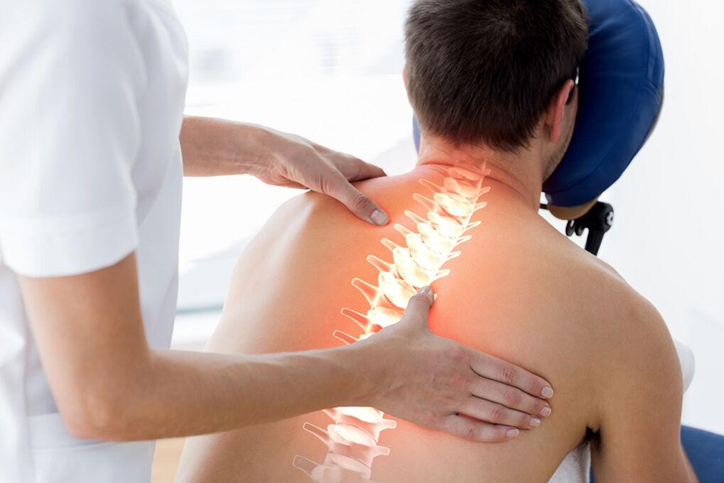
It is carried out by a neurologist or chiropractor after the therapist has ruled out pathology of the internal organs.
- The specialist finds out the main complaints, the time of onset, the development, the effect of the drug on pain intensity, rest, changes in the rhythm of life.
- Mandatory external examination is carried out when the patient is undressing - it is necessary to compare the condition and color of the skin on the symmetrical parts of the body, the tone of the tissues, the response to stimulidifferent: pain, touch, cold. or heat. Stress symptoms are identified, showing muscle tension and irritation of the tendons and their connective membranes - the fascia
- The nerve hammer will reveal the evenness and symmetry of the reflexes
- The neurologist records the volume of active (independent) and passive (performed by the doctor) movements in the joints, the ability to turn the head, the upper body without involving the lower part of the columnliving.
If necessary, send for additional testing
- Diagnostic thermal imaging
- ENMG (electroneuromyography): Radiographic imaging. To obtain the necessary information, it is performed in at least two projections - direct and lateral. The imaging will reveal the state of the bone tissue, the severity of the osteoporosis, the size and safety of the vertebral bodies, and will reveal the osteogenic cells. Damaged discs are determined by the width and uniformity of the disc fissures. Unevenness of the lower or upper body contour would raise suspicion of a Schmorl hernia. To clarify the nature of changes in the bony structure of the spine, computed tomography is recommended. The multi-cavity examination allows the creation of a three-dimensional model of the vertebrae. If necessary, to find out the condition of the soft tissues - muscles, ligaments, discs, MRI is prescribed.
It must be remembered that the results of the study must be compared with the complaints and changes found during the examination. Detecting signs of spondylolisthesis and even disc herniation without complaints does not require any serious measures.
Treatment of osteonecrosis of the spine
Eliminates acute manifestations of the disease
- The intense pain and strong muscle tension reinforce each other, not allowing the exacerbation to subside. Therefore, the first step is pain relief.
- Prescribing injectable non-steroidal anti-inflammatory drugs, muscle relaxants - muscle relaxants
- If these measures are not sufficient, blockade with analgesics and hormonal drugs is instituted
Reduce radio frequency
Should rest in bed for a few days
After the symptoms subside, it is necessary to initiate movement, gradually increasing the range of motion and the load. At this time, kneading and massaging activities are undesirable due to possible complications.
Osteochondrosis: treatment without exacerbations
When the patient's condition is stable, the usual lethargy persistsbone necrosis, treatment includes several components:
- Medicine. All anti-inflammatory pain relievers are the same in the form of tablets, capsules, and ointments. A specific drug is selected by the physician based on the patient's condition, lifestyle, comorbidities, predominance of one or another component of osteonecrosis. A course of B vitamins improves the conduction of impulses along the nerves, normalizes tissue nutrition. While maintaining muscle tone, the use of muscle relaxants should be continued. The drug relieves symptoms, improves mobility and performance. But they cannot completely stop the progression of the disease.
- Physical therapy. It is used to deliver medication directly to the painful area (electrophoresis), to warm it (paraffin irradiation, infrared irradiation). Exposure to therapeutic electric currents relaxes muscles, improves the functioning of nerve fibers. After a few sessions, the pain subsides and mobility is restored. Not prescribed for active inflammation
- Manual manipulation, massage, acupuncture, acupressure. Relieve spasms by stretching and relaxing the muscles. If during the massage process only affects the upper muscle layer, manual therapy will penetrate deeper, so the requirement for a specialist is higher. Make sure to do an MRI first to find out the anatomical features of a particular patient
- Spinal traction. The vertebrae move away from each other, the normal distance between them is restored, the compression of the nerves is reduced. The procedure has contraindications so only a doctor can prescribe it
- Physical therapy. The most effective treatment. The only word of warning is that it must be applied to life. Among the advantages - it provides activity, improves mood, increases tissue tone. The best methods are doing the exercises recommended by the doctor, the initial yoga poses, Pilates, swimming. They are performed smoothly, without sudden and traumatic movements, stretching the tissue, gradually increasing the amplitude
- Proper nutrition and giving up bad habits
- Adequate nutrition to the tissues, good condition of blood vessels, and adequate blood supply to the vertebrae and surrounding structures are measures to prevent the progression of osteonecrosis. Proper nutrition helps to normalize weight and reduce stress on the spine
Surgical treatment of spinal osteonecrosis.Modern clinics have a large arsenal of minimally invasive interventions:
- Treatment and diagnostic blockade
- Cut off the radio frequency aspect
- Cold plasma and laser nucleation
- Endoscopic removal of herniated disc
- Micro cutout
Radiofrequency thermal ablation of facet joints
Special needles are placed precisely on the side of the disc joints at the place where the median branch of the Lyushka nerve passes. The electrodes are inserted into the needle, the tip of the needle will heat up to 80 degrees in 90 seconds. This leads to blood clotting of the nerve. End of pain.
nucleation with cold plasma
Through a needle inserted into the disc, a special cold plasma electrode is inserted into the disc tissue. Endothelial pressure is reduced, the hernia (convex) is pulled inward.
Micro cutout
With a herniated disc, nearby nerve roots and blood vessels are compressed, causing extreme pain and internal dysfunction of the extremities. If conservative treatment is no longer effective, surgery for herniated disc is the only viable solution for many patients. The surgery is performed under anesthesia through a 2-3 cm long incision using microsurgery equipment and instruments. The duration of the operation is 45-60 minutes. Pain syndrome significantly decreased or completely disappeared immediately after surgery in 95% of patients. The next day the patient was able to walk and was soon discharged from the hospital.
Endoscopic removal of herniated disc:
Herniation or free-standing tumor is removed through the lateral discs. To place the tube, a 5 mm incision is made in the skin. The muscles, balance and ligaments are not damaged, they are pushed out using an increasing diameter retractor system. The surgery requires almost no blood and lasts only 40-50 minutes. Patients can return to their usual regimen after three weeks. The risk of complications is minimal.
Decompression and stabilization operations are performed when complications arise, large disc herniation, severe compression of spinal nerve roots and spinal cord. If there are signs of sudden loss of sensitivity, movement, pelvic dysfunction, the patient should be urgently referred to a neurosurgeon. The sooner the compression is removed, the more complete the recovery is, and the patient will quickly return to a normal life. In this case, surgical treatment is aimed at decompressing the compressed nerve structures and stabilizing the affected segment. This is a hemi- or laminectomy. Fixation is accomplished by a lens system, which, combined with a mid-body cage, provides 360-degree fusion. Endothelial stabilization of the vertebrae is widely used. There are several methods of transendothelial transplantation today. Microdiscectomy combined with intervertebral stabilization, especially in the elderly, can significantly increase long-term efficacy and reduce the likelihood of herniated disc recurrence.